

 First posted November 29, 2007 Last updated August 21, 2013
Children's Distal Radius Fractures
(also known as a "broken wrist")
![]()
Children's Distal radius fractures are some of the most common fractures (the medical term for "broken bone") in children, and they are very different from adult distal radius fractures.
What does a normal child's bones look like?
 |
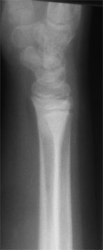 |
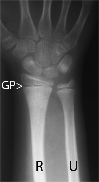
Normal child, about age 6, two views
PA view on the left, lateral view on the right
In the two xrays above, the image to the left is looking at the forearm and hand from the back side of the hand. The image to the right is looking at the hand and forearm from the side. The radius is the forearm bone on the thumb side, on the left xray above, it is the bone on the left, marked R. The second bone of the forearm is called the ulna. In the left xray above, the ulna is on the right side of the image, marked U. The image to the right is looking at the hand and forearm from the side. The radius and ulna images are on top of each other, making them hard to see. The black line across the bones, very near the end, is the growth plate (marked GP), how the arm gets longer.
Distal radius fractures are generally caused by a fall on an outstretched hand. The fracture is almost always within an inch of the wrist joint, and may extend into the joint.
![]()
Types of Children's Distal Radius Fractures
There are two general types of children's distal radius fractures: (1) fractures that involve the growth plate and (2) fractures that do not involve the growth plate. Any of these fractures can be either displaced (out of normal alignment) or nondisplaced. Fractures in (2) can generally be classified as either buckle fractures or shaft fractures. Each of these fractures is unique in what has been injured and how they are treated.
![]()
Treatment of Distal Radius Fractures
The treatment depends on the types of fractures.
Buckle fractures that are not angulated are generally splinted in place for a few days to a week, then placed in a cast, for a total length of immobilization of about 6 weeks. Buckle fractures that are angulated more than about 5 degrees are generally reduced. This involved placing a numbing shot in the arm and bending the bones back towards straight. Due to the springyness of children's bones, they usually cannot be make to be completely straight. Luckily, due to the ability of a child's bones to remodel (grow straight on their own), the bones do not need to be made straight by the orthopedist.
Displaced fractures
![]()
What Can I expect While My Child Is Healing?
Children usually do not have as much pain as adults do with similar fractures. Aspirin or Tylenol is usually all that is requires. Sometimes Tylenol with codeine is given. Elevation and rest is best for the first day or so. You can add ice if you want. I think that you will find, as I have over 23 years of treating children's fractures, that you will be surprised by how quickly your child returns to being very active. Most patients are back to close to normal activity within one to three days. After the first day or so, the challenge is usually restricting the child's behaviour, not managing the pain.
![]()
What Can I Expect After It Has Healed?
Most children's forearm fractures heal very well. Buckle fractures are the quickest to heal and within two months you may not be even able to see the fracture on the xrays, much less by looking at the arm. Displaced fractures may show a slight deformity when you look at the arm, but as the child grows and the bone remodels, the deformity will disappear in almost all cases (less than 5 in my 23 years have not disappeared). Growth plate injuries need to be followed for about 3 to 6 months, to be sure that the bone grows normally. Growth plate injuries that lead to deformity are serious, but also are luckily so rare that I have not yet seen one in my own patients in my entire career.
![]()
Dr. Nelson has a particular interest in distal radius fractures. He has chaired several international courses exclusively on distal radius fractures, and has lectured on them in the United States, England, France, Italy, Argentina, Brazil, Turkey, and Japan. He has invented one of the external fixatorsand one of the internal fixation plates that are used to treat these fractures in adults. These devices are sold throughout the world. He is actively involved with anatomy research, developing newer methods of distal radius fracture treatment and with teaching surgeons from around the world about how to treat distal radius fractures. He is the Webmaster of a site, eRadius.com, devoted exclusively to distal radius fractures. (eRadius is designed to teach experienced orthopedic and trauma surgeons; it is not designed for patients.)
![]()