
Advanced Dupuytren's Page
Palmar fascia
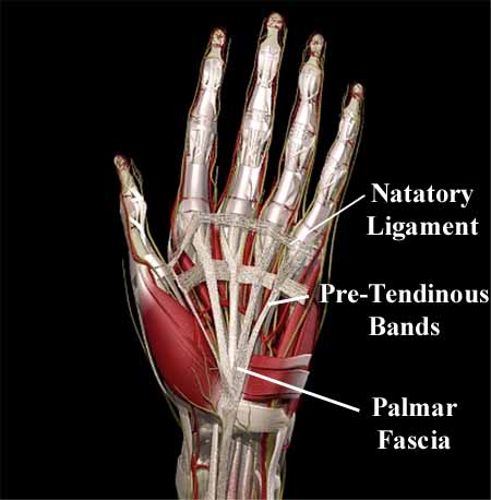
The fascial structures in the palm comprise distinct longitudinal, transverse and vertical (i.e. at right angles to the plane of the palm) components (these terms are clinical rather than relative to the anatomical position).
The longitudinal fibers are the distal continuations of the fibers of palmaris longus. Distal to the distal palmar skin crease, the longitudinal fibers separate into 3 layers.
The intermediate longitudinal fibers pass deeply into the tissues of the webs spiraling behind the neurovascular bundles to reach the digits (lateral digital sheet).
Note that there is a continuation of the natatory ligament that extends across the first webspace into the thumb.
All of these tissues can become involved in Duputyren's disease.
On a deeper plane in the mid palm, deep to the transverse fibers of the palmar fascia, a series of vertical fibrous septae separate the flexor tendon sheaths to each digital ray.
The fascial structures of the palm are a complex three dimensional lattice stabilizing the distal palmar skin while allowing limited movement. The fascia protects the underlying tendons, nerves, and blood vessels from receiving the external forces applied to the palm during frictional activities. This type of fascial tissue is markedly absent on the dorsum of the hand where the skin is loose and highly mobile.
The cords of contracture in Dupuytren’s disease tend to follow anatomical ligamentous pathways, and knowledge of the 3 layers of fascia makes surgery more precise.
At the mid-palmar crease the transverse fibers of the palmar aponeurosis lie superficial to the neurovascular bundles and therefore represent a safe surgical landmark.