
Tennis Elbow Histology
The study of our bodies at the microscopic level is called histology. Here are three stages of injury and healing that may help you to understand tennis elbow and therefore be better able to direct your own care.
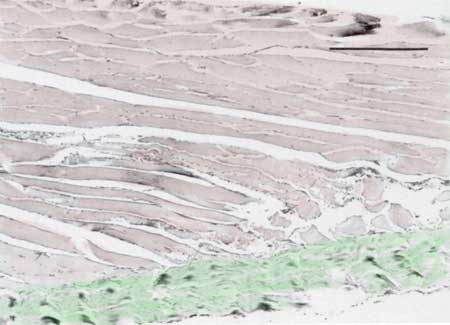
This is a microscopic view of a muscle (artificially colored pink) and its insertion into a tendonous tissue such as Sharpie's fibers (artificially colored green).
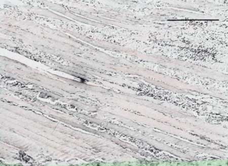
This is a microscopic view of a muscle (artificially colored pink) and its insertion into a tendonous tissue such as Sharpie's fibers (artificially colored green), 48 hours after a muscle tear. Compare this view to the one above. Notice the large number of slightly blue cells, which are the inflammatory cells.
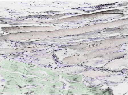
This is a microscopic view of a muscle (artificially colored pink) and its insertion into a tendonous tissue such as Sharpie's fibers (artificially colored green), seven days after an injury. Notice that there are fewer inflammatory cells (artificially colored blue).
| Would you like to search the medical library of the National Library of Medicine for scientific papers on this topic? Just click on the Pub - Med image: | |
| Remember the admonition from the Patient Education Links Page: the Internet has a lot of information, much of it incorrect. I have reviewed the sites that I have linked to, and have only linked to sites when I personally know the surgeon who posted it, or am a member of the organization that posted it. However, I may not agree with all that is on that site, and it may have changed since I reviewed it. If any of the information is not consistent with what I have told you, please download the material and bring it in. | |